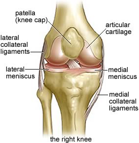
For chiropractors, we have to look at a patients complete anatomy even someones knee to really get down to the cause of your pain. The skeletal structure of the knee is formed from three different bones – the femur (thigh bone), tibia (shin bone) and patella (knee cap). The lower end of the femur connects with the tibia and patella at the knee. Here, the femur expands into two protuberances known as the medial and lateral condyles, which meet with both the medial and lateral condyles of the tibia and two articular facets of the patella.
As the femur inclines inward from the hip, the knee joints are closer to the midline of the body than the hips. This inclination tends to be greater in females because of their wider pelvis. Unlike other joints in the body, which are mostly formed by the intersection of two bones, the knee comprises three different connections: two tibiofemoral hinge joints and the planar (sliding) patellofemoral joint. The tibiofemoral joints are formed where the lateral and medial condyles of the femur meet with the lateral and medial condyles of the tibia, respectively. Two articular menisci (fibrocartilage discs), one lateral and one medial, also form part of these joints.
 These act to fill the spaces between the bones and help with the circulation of synovial fluid. The patellofemoral joint is formed by the convergence of the patellar surface on the front of the femur with the back of the patella. Movements permitted at the knee joint include flexion (bending), extension (straightening), a small amount of medial (inward) rotation and some lateral (outward) rotation of the bent leg.
The joint is supported by a series of muscle tendons, ligaments and some capsular fibers. If you have knee pain contact us at Chicago Chiropractic, chiropractors can help even in this area! Tendons from the quadriceps femoris muscle stretch across the front of the patella, forming both the lateral and medial patellar retinacula and then continue down to the tibia as the patellar ligament. These structures provide the front of the patellofemoral joint with a great deal of strength. Behind the patellar ligament a fatty cushion (the intrapatellar fat pad) separates the ligament from the synovial membrane. Other important supporting ligaments at the knee include two popliteal ligaments (the oblique and arcuate, which strengthen the connections between the tibia and femur), two collateral ligaments (the tibial and fibular, which connect the femur to the tibia and fibula) and two cruciate ligaments (the anterior and posterior) that stretch across the inside of the knee joint, crossing as they do so (hence the name cruciate which means ‘cross-shaped‘).
The cruciate ligaments are well-known to many sports fans, as both (especially the anterior) can become badly damaged by any strong twisting of the knee following a fall. Three bursae (fluid-filled sacs) are found at the knee joint, which act to reduce friction where connected parts of the joint move against each other. As a chiropractor we have to take this very seriously. These comprise the prepatellar bursa between the patella and skin of the knee, the intrapatellar bursa between the top of the tibia and patellar ligament and the suprapatellar bursa between the bottom of the femur and quadriceps femoris muscle.
